An ultrasound is not just a photoshoot but an essential diagnostic tool. It provides your doctor with lots of valuable information. They monitor your baby’s growth, detect abnormalities, predict your due date and determine the sex and number of babies you are having. Conducted during pregnancy, these ultrasounds show the position of the placenta, which helps your gynecologists take the best decisions regarding your baby’ health. Consult an experienced and board-certified gynecologist at the Century Medical and Dental Center to learn how ultrasounds work and how they provide doctors the information necessary to make a diagnosis if they detect any problems.
Also known as a sonogram, an ultrasound is typically performed by an ultrasound technician or sometimes by an OBGYN. It uses sound waves to create an image of the organs inside your body. Ultrasounds during pregnancy help doctors diagnose many diseases and conditions, even those not related to pregnancy. These sound waves are not harmful to you or your baby.
Mothers-to-be look forward to ultrasounds during pregnancy more than any other appointment. It gives them a sneak peek of their bundle of joy while the specialist looks for specific growth and development marks. Some women also want to know about their baby’s sex and wait impatiently for their 20-week ultrasound. Keeping your ultrasound appointments is necessary to learn about the baby’s growth and determine if other tests need to be done to know about your baby’s health.
This article covers why and how ultrasounds are done during pregnancy. It also discusses how many ultrasounds to expect while you are pregnant, and what the OBGYN is looking for. Most pregnant women get only two ultrasounds, one at the beginning of pregnancy and one about halfway through. Some women may have three or more ultrasounds depending on several factors.
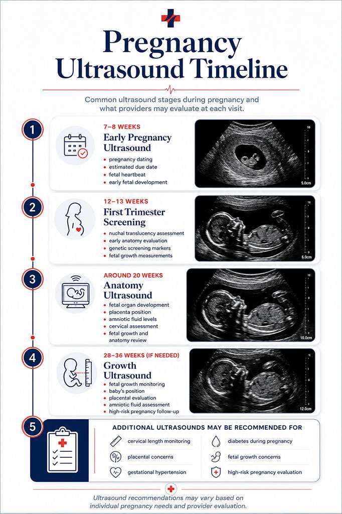
The first ultrasound is called the dating or viability ultrasound. It is typically done between 7 and 8 weeks to verify your due date, look for a fetal heartbeat, and measure the baby’s length from crown to rump. At this ultrasound, you will also learn whether you are having one baby, twins, or even more. At these sessions, you may even get to see or hear the baby’s heartbeat.
If you have irregular periods or did not have a period after coming off birth control, this ultrasound will help determine an accurate due date. The due date is crucial as it allows the doctor to keep an eye on fetal development and track it every month.
When you are 7 to 8 weeks pregnant, your fetus is only about two centimeters long. To get a close view of your uterus and the developing fetus, a dating transvaginal ultrasound is performed. It means the ultrasound is done internally through the vagina. A transvaginal ultrasound can be a little uncomfortable, but it is not painful. Many women reported it being less invasive than the speculum used during a gynecological exam.
The OBGYN or the ultrasound technician will gently insert an ultrasound wand inside your vagina. The transvaginal ultrasound wand is also called a transducer. It’s about three centimeters around, a little larger than a tampon. It will be covered by a condom and lubricant. The wand will not reach your cervix and take images of your uterus. It is perfectly safe for your baby.
The doctor may ask you to come for the first ultrasound with a full bladder. A fuller bladder enables the doctor to put your uterus in a better position and get clean images.
What the doctor looks for at the first ultrasound
If you are opting for prenatal genetic testing, you will have your next ultrasound at 12 to 13 weeks gestation. This ultrasound is also called nuchal translucency screening. It is offered to every mother-to-be and is covered by most insurance plans. The genetic screening ultrasound is optional.
During this ultrasound, the doctor will look for indicators of chromosomal disorders. Chromosomal disorders mean that the baby received an extra chromosome at conception and could have moderate to extreme physical or mental challenges.
These disorders include:
This ultrasound is an anatomical scan. The doctor will look to see if all four limbs are present. They will also look for basic structures in the brain, the stomach, the bladder, the nasal bone, and something called nuchal translucency. Nuchal translucency is a fluid sack at the back of the baby’s neck filled with lymphatic fluid. There are correlations between the size of that sack of fluid and the likelihood that the fetus could be affected by a major chromosomal disorder.
After the ultrasound is complete, the doctor will interpret the results and share its information with you. If needed, the doctor may recommend you consult a genetic counselor who could recommend having additional tests done to verify the ultrasound results. You must understand that ultrasound screenings for other genetic disorders or anatomic abnormalities become more accurate as the pregnancy advances.
The decision to go for a genetic screening ultrasound is yours only and depends on the doctor’s recommendation to an extent. There are some essential questions you must ask yourself when thinking about genetic screening. They include:
Going for a genetic screening ultrasound at this time is up to you. Some women prefer to have as much information as they can gather early in their pregnancy, while others do not. Discuss the pros and cons of genetics screening with your doctor to know if it would help you and it is a necessity in your case.
It is the ultrasound that parents-to-be look forward to the most. The full anatomy ultrasound is typically performed at about 20 weeks or 5 months. As evident from its name, this ultrasound looks at all the baby’s organ systems to make sure they are present. It checks if they are a normal size and shape, and are in the right location.
The full anatomy scan is a transabdominal ultrasound. It uses a transducer that looks a lot like a store checkout scanner. The ultrasound technician will put warm ultrasound gel on your stomach and then slide the transducer in the gel around your stomach. The gel helps the sound waves travel through your skin.
The doctors advise you to come for an ultrasound with a relatively full bladder as it makes it easy for the technician to get better images of the baby.
There are many things to look for in a complete anatomy scan ultrasound. It takes around 45 minutes if the baby cooperates. If you have a squirmy baby who is not so camera-friendly, it could take a few hours to get all the images the doctor needs to check things out. The doctors know all the ways and tricks that encourage the baby to change positions. They include asking you to lie down on one side then the other and emptying your bladder or filing it or maybe even walking around. The doctors do all it takes to take images to track your baby’s growth and development.
During the full-anatomy 20 week ultrasound, you can find out if your baby is male or female. If you want the baby’s sex to be a surprise, let your technician know this so that he or she does not let it slip accidentally. When the scan is complete, the clinic can even send you a link to view some fun photos of your baby.
The ultrasound technician will capture several images and measurements. They include:
Once the ultrasound technician has captured all the images and measurements, the OBGYN will take a good look at them to check for abnormalities such as congenital heart defects or cleft lip or palate. They will discuss their conclusions with you and help you understand what you are looking at in the different images and what they mean. If everything looks normal and there are no problems, and things continue well, you will be holding your baby in your arms within a short period. In the meantime, you can enjoy those 2D or 3D photos of your baby.
Sometimes women need additional ultrasounds during pregnancy. The OBGYN may ask you to come in for additional ultrasounds to look for certain elements. They include:
If your cervix is shorter than expected, you may need to have your cervix checked regularly to be sure it stays closed to maintain your pregnancy. If the cervix continues to shorten or thin, you may need a cerclage to help strengthen it till it is time to deliver the baby. Cervical length ultrasounds occur at 16, 18, 20, and 22 weeks and are done transvaginally.
If your placenta is too small, it is in an abnormal location or if it is an abnormal shape, the doctor will need to monitor it and the growth of your baby with regular ultrasounds. The placenta is responsible for passing blood and nutrients to your baby, so it must grow correctly.
You will require growth ultrasounds if you have:
Sometimes growth ultrasounds are required to check that the baby’s growth is continuing along the growth curve. They are done at 28, 32, and 36 weeks. The doctor checks out if your baby is growing as expected by measuring your fundal height. Fundal height is the number of centimeters from the pubic bone to the top of your uterus. This measurement typically increases by about 1 cm each week. If your uterus has not grown appropriately in the last month, the OBGYN will determine that your baby is also not growing well and will want to perform monthly growth ultrasounds.
These ultrasounds take less time than the full basic anatomy ultrasound because there are fewer measurements required. The ultrasound technician will measure the baby’s head circumference, bi-parietal diameter, abdominal circumference, and femur length.
The OBGYN wants to see if your baby is staying on its growth curve. The doctor uses the measurements to estimate the baby’s weight. A large or extra-large baby is not very concerning, but an extra small baby or baby which is not growing according to the growth curve could mean that the baby is not getting sufficient nourishment through the placenta. Such babies may need to be delivered early.
2D, 3D & 4D ultrasounds
Many ultrasound centers offer to tell you your baby’s sex early on or give you keepsake 3D or 4D images. They are not necessary and are rarely covered by insurance. You will find out everything you need to know during your appointments at the clinic, which are covered by the insurance. A clinic is the best place to go for ultrasound as you will have access to a trained and qualified physician who can interpret the images accurately. It will not only add to your joy but also help the gynecologist keep an eye on your baby’s growth and development.
Ultrasounds during pregnancy are a fascinating way to get a glimpse of your developing baby. Visit your gynecologist at Century Medical and Dental Care to know more about genetics screening and full anatomy scans and how they can help you welcome a healthy baby into this world. The experienced and trained doctors in NY check for how long you are in pregnancy and determine if other tests need to be done to track the baby’s progress. They do their very best to keep you safe and informed and ensure the best outcome for your pregnancy.
SHARE THIS POST Page Updated on May 12, 2026 by Dr. Dvorkina (Primary Care Doctor) of Century Medical & Dental CenterCentury Medical and Dental Center is an accredited healthcare facility in NY that operates in accordance with Article 28, a public health law. This law regulates and recognizes accreditation for public healthcare facilities, ensuring they are licensed and operated correctly. By undergoing the Article 28 process and achieving accreditation, Century Medical and Dental Center demonstrates its commitment to meeting the highest standards of care.
As a multidisciplinary medical center, we have highly qualified doctors, nurses, and support staff who are working hard to provide the best medical care to patients in Manhattan, NY, Brooklyn, NY, and Bronx, NY including Brooklyn Heights, Dumbo, Prospect Heights, Park Slope, Clinton Hill, Boerum Hill, Red Hook, Harlem, Gravesneck, Flatbush, and Bedford-Stuyvesant.